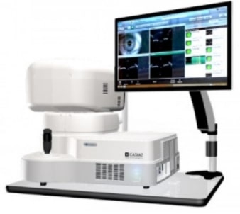
CASIA2
Advanced Imaging Cornea and lens are shown in one image [Deeper imaging]
With CASIA2, the light source of coherency functions is improved, and higher sensibility toward depth is realized compared to our former model. By using this new technology, it is possible to measure from anterior cornea to posterior lens with one shot. The scanning depth is approximately 13 mm. CASIA2 explores new possibilities in anterior segment analysis.
[Wider imaging]
Capturing images around the angle is possible in corneal topography mode. As with corneal shape analysis, it is possible to extract and analyze the angle and observe the IOL, which enables testing without switching measuring modes.
[Clearer imaging]
By scanning 16 images simultaneously, clearer images are obtained.
Application for cataract surgery CASIA IOL Cataract Surgery(CICS)
The testing application for cataract surgery, CICS, is installed in the CASIA2, which effectively supports cataract surgery. There are two types in CICS: "Pre-op testing" and "Post-op testing". To use their functions, capture the image within each testing protocol.
Fulfilling analysis functions
- Trend analysis
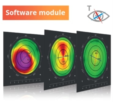
In addition to the color code map showing parameter changes of each corneal shape, the simplicity of the graph means information is instinctively easy to grasp. Hence, this analysis is useful for observing the keratoconus.
- Lens shape analysis
While capturing the anterior cornea to the posterior lens, it is possible to measure the corneal curvature, thickness, and tilt of the anterior/posterior lens.


