IHC
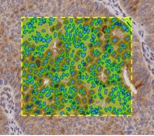
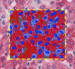
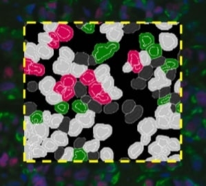
The Aperio Image Analysis IHC menu provides a broad range of solutions for quantification of single and multiplex tissue staining. Flexible algorithms are easily optimized for diverse brightfield chromogens, enabling you to customize the analysis to match your unique research needs. Accurately identify protein biomarker expression at the tissue, cellular or subcellular level.
Products
Aperio Membrane Algorithm
Identify staining specific to the cell membrane, while eliminating non-specific staining in other subcellular compartments.
Aperio Nuclear Algorithm
Quantify the intensity and amount of staining in cell nuclei, while including only cells of interest based on nuclear morphology and size.
Aperio Cytoplasm Algorithm
Measure staining located within the cell cytoplasm, while excluding non-specific staining in the membrane or nucleus.
Aperio Rare Event Detection Algorithm
Detect and count objects of interest, such as circulating tumor cells (CTC) or micrometastases, based on color and size.
Aperio Microvessel Analysis Algorithm
Automatically detect and measure microvessls, and quantify overall density of vasculature in tissue.
Aperio Color Deconvolution Algorithm
Separate color channels in multiplex stained images, to identify location, area and intensity of each stain.
Aperio Colocalization Algorithm
Determine location of multiple stains, and automatically quantify colocalization of stains across tissue.


