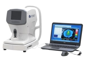
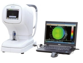
TMS-4N
High usability and reliability
With just a few touches on the main screen, it is easy to access the map analysis function. In addition to the keratoconus screening function and Fourier Analysis function to quantify astigmatism, several reliable analysis tools provide accurate diagnostic information.
Auto alignment with 3D eye tracking method
Simply adjusting the alignment light on the first ring starts automatic measuring, making the TMS-4N accurate for even the less-experienced examiner. As the TMS-4N produces consistently clear images through auto-shot alignment, the risk of incorrect color-code maps resulting diminishes significantly.
Several analysis tools
In addition to the Klyce / Maeda method, keratoconus evaluation uses the Smolek / Klyce method of neural network classification. Keratoconus (even a mild case), is a contraindication for refractive surgery, and this evaluation is useful for screening tests.
With the Fourier analysis function, a normal color code map is separated into a spherical component of the cornea, a regular astigmatism component, an asymmetry component, and higher order irregularity to evaluate a more detailed corneal shape. This function helps evaluate an irregular astigmatism component in corneal diseases and pre / post refractive surgery.
The power difference map displays changes in corneal curvature. It is useful for evaluating a change in corneal shape after cataract surgery and centering evaluation after refractive surgery such as orthokeratology.
Axis registration method
From the captured image, it is possible to check the marking and the degree of marking can be measured.
By using the obtained value, the fixation axis of the Toric IOL can be confirmed with higher accuracy
14 kinds of corneal shape indexes
The TMS-4N installs 14 kinds of corneal shape indexes such as a corneal eccentricity index (CEI) which converts the flattening rate of the cornea. Import data from our former models; TMS-1, TMS-2, TMS-2N and TMS-4N to display maps and analyze data.


