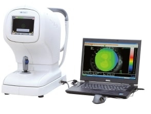
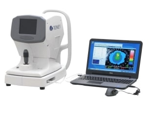
TMS-5
A Scheimpflug camera which enables measurement in bright light
In addition to the functions of the former TMS series which reflect mire ring images on the cornea, the TMS-5 can measure an anterior segment image (Scheimpflug image), by rotating and radiating slit light. The slit mode of the Scheimpflug camera is using a slit cone method, so it is less likely to be influenced by external light, which accommodates measurements in bright light.
* An attachable hood may be required according to the circumstances in the testing environment.
Anterior corneal map (merged) screen to improve analysis ratio
In higher order irregular astigmatism, with a ring topo mode and slit mode measurement, the Scheimpflug image is merged in ring topo data to provide an "anterior corneal merged map". The Scheimpflug image covers the area that ring topo data cannot obtain, and it becomes possible to display a merged map.
Instant capture and easy testing
A touch panel is installed in the LCD screen of the measurement head, so basic actions can be completed via the main body. The joy stick moves up and down and the angle of the screen can be adjusted by the examiner.
Analyze all data of the anterior/posterior segment of the cornea by ring topography mode and slit mode
- 4 map screen
If the Scheimpflug image is captured after ring topo data measurement by ring cone, it is possible to create an anterior corneal merged map, anterior / posterior corneal elevation map and pachymetry map.
- Slit calculation screen
On the slit calculation screen, it is possible to measure anterior chamber depth (ACD) and central corneal thickness (CCT) in addition to observing the anterior segment.
[Map screen]
- 1. Anterior corneal elevation map
- 2. Posterior corneal elevation ma
- 3. Anterior corneal merged map
- 4. Pachymetry map
- 5. CCT : central corneal thickness (distance between CCT-F and CCT-B)
ACD [Epi.] : anterior chamber depth including CCT (distance between CCT-F and ACD-L)
ACD [Endo.] : anterior chamber depth excluding CCT (distance between CCT-B and ACD-L)
*AR1, AR2 : angle recess 1, angle recess 2
Analyze anterior segment 3D imaging with Scheimpflug imaging
- Ectasia screening
On the ectasia screening screen, the anterior and posterior corneal shapes are both considered to show the screening results of ectasia patterns including keratoconus.
- IOL power calculation software "OKULIX"
The IOL power calculation software "OKULIX"is available to confirm the IOL power in cataract surgery after a LASIK operation.


