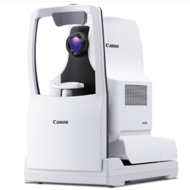
Xephilio OCT-S1
SUPERIOR PENETRATION OF DENSE OBJECTS
Revolutionary swept source technology allows you to capture wide-field images of up to 23 mm in a single scan. Xephilio OCT-S1 enables superior penetration of dense objects and provides outstanding tomographic images.

WIDE FIELD SWEPT SOURCE OCT IN ONE SINGLE CAPTURE
With Xephilio OCT-S1 Canon introduces revolutionary swept source technology allowing you to capture wide-field images of up to 23 mm in a single scan. Xephilio OCT-S1 enables superior penetration of dense objects and provides outstanding tomographic images. Experience a new quality of OCTA images in a single scan with greatly reduced noise, increased detail and improved visibility within just seconds.
AI-POWERED PERFORMANCE
AI helps you save time and improve imaging
Canon’s Intelligent Denoise, the system’s Deep Learning AI technology, offers a new quality of OCTA images based on individual scans – without the need to acquire and merge multiple images. The revolutionary technology delivers images with greatly reduced image noise, increased detail and improved visibility within just seconds.
FASTER
100,000 A-scans per second combined with invisible 1,060 nm wavelength provide ultra-fast swept source technology maximizing data quantity of the patient’s eye while reducing acquisition time. Invisible scan lines ensure better patient collaboration and reduce the impact of patient eye movements.
DEEPER
Canon’s deep scanning swept source technology allows better penetration of cataracts, hemorrhages, blood vessels and sclera and at the same time optimizes capture of retinal and choroidal data – all in a single shot. With Xephilio OCT-S1 vitreous body and choroid appear in the same image with superior image quality providing more information for better patient care.
WIDER
With a single capture the swept source Xephilio OCT-S1 shows a large wide-field OCT image as wide as 23 x 20 mm, which can be very beneficial for retina thickness observation of retinal detachment or retinitis pigmentosa. Mosaic imaging allows you to create an incredible 31 x 27 mm wide field OCT image.

23 mm wide-field OCT B-scan image

31 x 27 mm OCTA mosaic image
Connectivity with Ophthalmic Software Platform RX
Multimodality platform for all Canon retinal cameras and OCTs. The Ophthalmic Software Platform RX is designed for seamless integration and connectivity with patient management systems.
OUTSTANDING IMAGING IS YOUR BEST FRIEND
Canon’s recognized optical expertise enables the Xephilio OCT-S1 to offer superb image quality with minimal scatter. The swept source technology results in enhanced penetration further into the deeper tissue structures such as the choroid and even the sclera. Imaging depths of up to 5.3 mm allows for detailed visualization of the vitreous body and choroid in a single scan while the high scanning speed of 100,000 A-scans/s reduces examination time and offers very high resolution scans.

WIDER AND DEEPER
With Xephilio OCT-S1 wide-field images of up to 23 mm width can be acquired in just one scan, equaling an 80° viewing angle. The 5.3 mm depth allows for visualization of the vitreous body and choroid in a single scan with superior image quality.
SINGLE CAPTURE WIDE-FIELD OCT
Xephilio OCT-S1 provides wide-field imaging of up to 23 ×20 mm width with just one scan. Mosaic imaging allows you to create a wide-field OCT image of approximately up to 31 x 27 mm with just 4 or 5 images.

En-face Sattler’s layer, En-face Haller’s layer and Mosaic en-face image showing choroidal vortex veins.
EASY AND QUICK OPERATION
The Xephilio OCT-S1 utilizes a joystick for initial anterior alignment, but the operation is also aided by several automated functions. It has built-in SLO for real-time retinal tracking and accurate follow-ups.

The system’s joystick provides easy, quick operation combined with pin-point precision.
The built-in optimization function automatically takes care of alignment, focus and C-gate.

The wide-field SLO images acquired with Xephilio OCT-S1 allow for superior observation.
VERSATILE REPORTING POSSIBILITIES
Xephilio OCT-S1 provides you with a comprehensive range of reporting tools. Thanks to its extensive DICOM and EMR capability, results can be stored, shared and analyzed as needed in your daily practice.

3D reporting

OCT-A reporting
OUTSTANDING IMAGING.
With Xephilio OCT-S1 Canon introduces revolutionary swept source technology allowing you to capture wide-field images of up to 23 mm in a single scan. Xephilio OCT-S1 enables superior penetration of dense objects and provides outstanding tomographic images.

Vogt–Koyanagi–Harada disease. Courtesy of Kyushu University Hospital, Japan
VISUALIZE THE MICROVASCULATURE OF THE RETINA WITH OCT ANGIOGRAPHY
OCT angiography is a sophisticated technology that detects the movement of red blood cells in the retinal vasculature and allows you to visualize tiny vessels in detail.

Non-invasive examination, results within seconds
OCT Angio does not require fluorescein injection or pupil dilation, and the examination takes only seconds. SLO-based real-time tracking minimizes artefacts. Sophisticated image post-processing with 3D projection artefact removal enables excellent image quality.

Angio Expert with freely selectable layers
With OCT angiography even the smallest blood vessels can be observed in 2D and 3D. With Canon’s OCT Angio software, you can freely select layers to create the preferred image. Layers can be defined based on automatic segmentation or as a custom offset.

Analysis and reporting tools
Canon Medical’s Angio Expert software provides a comprehensive set of manual and automated analyses.

Automated area analysis and measurement
With a simple click on a non-perfused area or the foveal avascular zone, the target area is automatically detected, analyzed and displayed. If needed, users can change the automatically drawn borders or trace the area completely manually.
HIGH DENSITY AND SINGLE CAPTURE
The Xephilio OCT-S1 offers an enormous diversity of scan areas and scan densities for OCT Angiography examinations. While scan areas range from small (3 x 3 ~ 8 x 8 mm) to super large (23 x 20 mm), a high scan density of up to 928 x 807 pixels allows for visualization of small vessels at the same time.
Always the right angle
With Angio Expert, you can choose the optimal scan density for any viewing angle. The system provides various square and rectangular formats from 3 x 3 mm to 23 x 20 mm.

Single capture wide-field imaging
Single scan wide-field imaging enables OCT Angiography of up to 23 mm width.This allows wide-spread non-perfused areas to be visualized which is useful in diagnosing diabetic retinopathy and retinal vein occlusion. At the same time, a single high-density OCTA scan visualizes even small capillaries.

Panoramic imaging
The optional Mosaic software allows you to create ultra-wide OCTA images of approximately up to 31 x 27 mm.

Central retinal vein occlusion. Courtesy of Prof Staurenghi, Sacco University, Italy
CLINICAL CASES
Xephilio OCT-S1 enables superior penetration of dense objects and provides outstanding tomographic images.
RETINA
image C

image E

image A

C: Post Treatment Single-scan Wide-Field OCT Angiogram
A: Ultra Wide-Field Fundus Fluorescein Angiogram (UWF-FFA)
E: Wide-Field OCT Retinal Thickness Map
Courtesy of Prof. Paulo E. Stanga, Consultant Ophthalmologist & Vitreoretinal Surgeon, London Vision Clinic – Retina Lead, UK
BRANCH RETINAL ARTERY OCCLUSION


CHRONIC CENTRAL RETINAL VEIN OCCLUSION


RHEGMATOGENOUS RETINAL DETACHMENT


CENTRAL SEROUS CHORIORETINOPATHY


ANTERIOR SEGMENT OCT*
Xephilio OCT-S1 allows you not only to visualize the microvasculature of the retina, but also of the conjunctiva. The anterior segment can be observed without the need for any additional lens attachments.

En-face OCT and OCTA
* Anterior segment OCT is currently intended for research purposes only and must not be used for patient diagnoses.
INTELLIGENT DENOISE

Original image (w/o intelligent denoise)

Image with intelligent denoise
SINGLE SCAN WIDE FIELD OCT ANGIOGRAPHY
Post treatment single scan wide field OCT Angiogram. Perfused and non-perfused retinal areas are closely defined without the need of intravenous dye. Note that the residual fibrosed superior mid-peripheral NVE remains perfused and the residual minimally fibrosed temporal mid-peripheral NVE is perfused.


